
Parsley's Story
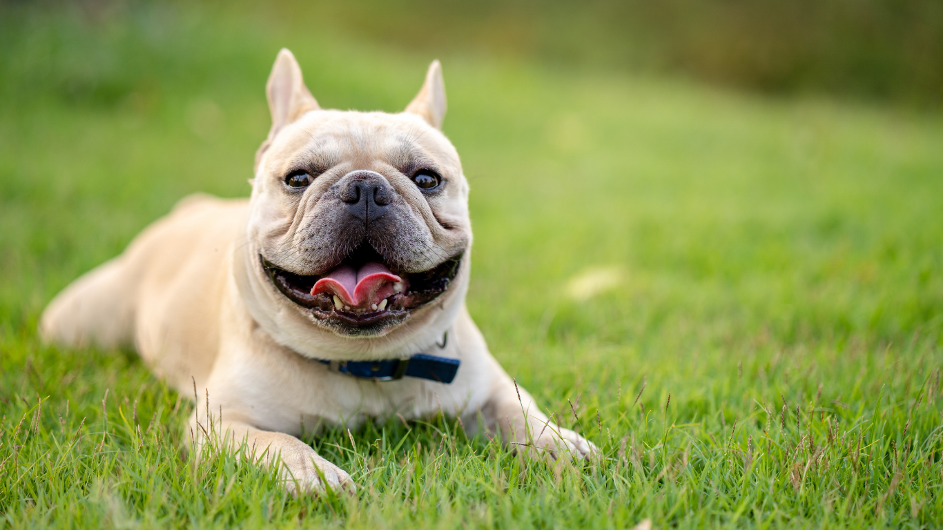
A French Bulldog like Parsley, whose years of chronic diarrhea were finally explained by an abdominal ultrasound.
Parsley’s primary vet said, "I was 50/50 she'd ever get better."
Years of Tummy Trouble
For most of her eight years, Parsley the French Bulldog had a gastrointestinal tract that never quite cooperated. Her family knew the pattern well: stress would hit, and so would the diarrhea. They managed it, year after year, the way loving families do: adjusting, worrying, hoping the next thing would be the thing that worked.
Then came the week that scared everyone. The symptoms turned severe and relentless. This wasn't the old familiar problem. Something had changed, and Parsley's family and veterinarian needed to know what.
The Search for Answers
Her veterinary team had already done thorough detective work: bloodwork, fecal testing, specialized gastrointestinal panels. Every piece of information mattered, but the complete picture was still just out of reach. To see what the other tests couldn't show, Parsley’s veterinarian called us for a complete abdominal ultrasound.
What the Ultrasound Revealed
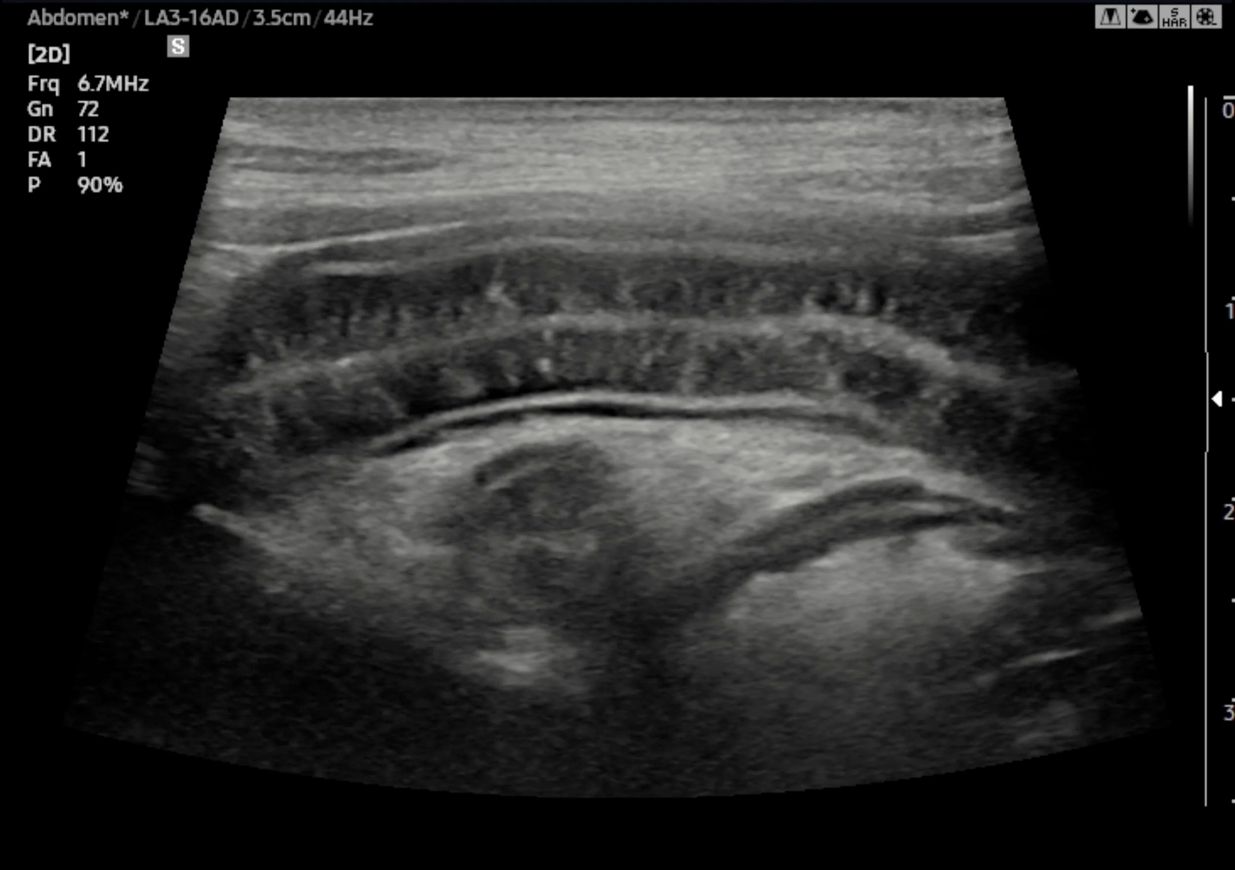
A close-up of Parsley's small intestine. The bright vertical streaks in the intestinal wall are swollen lymphatic channels, the telltale sign of lymphangiectasia.
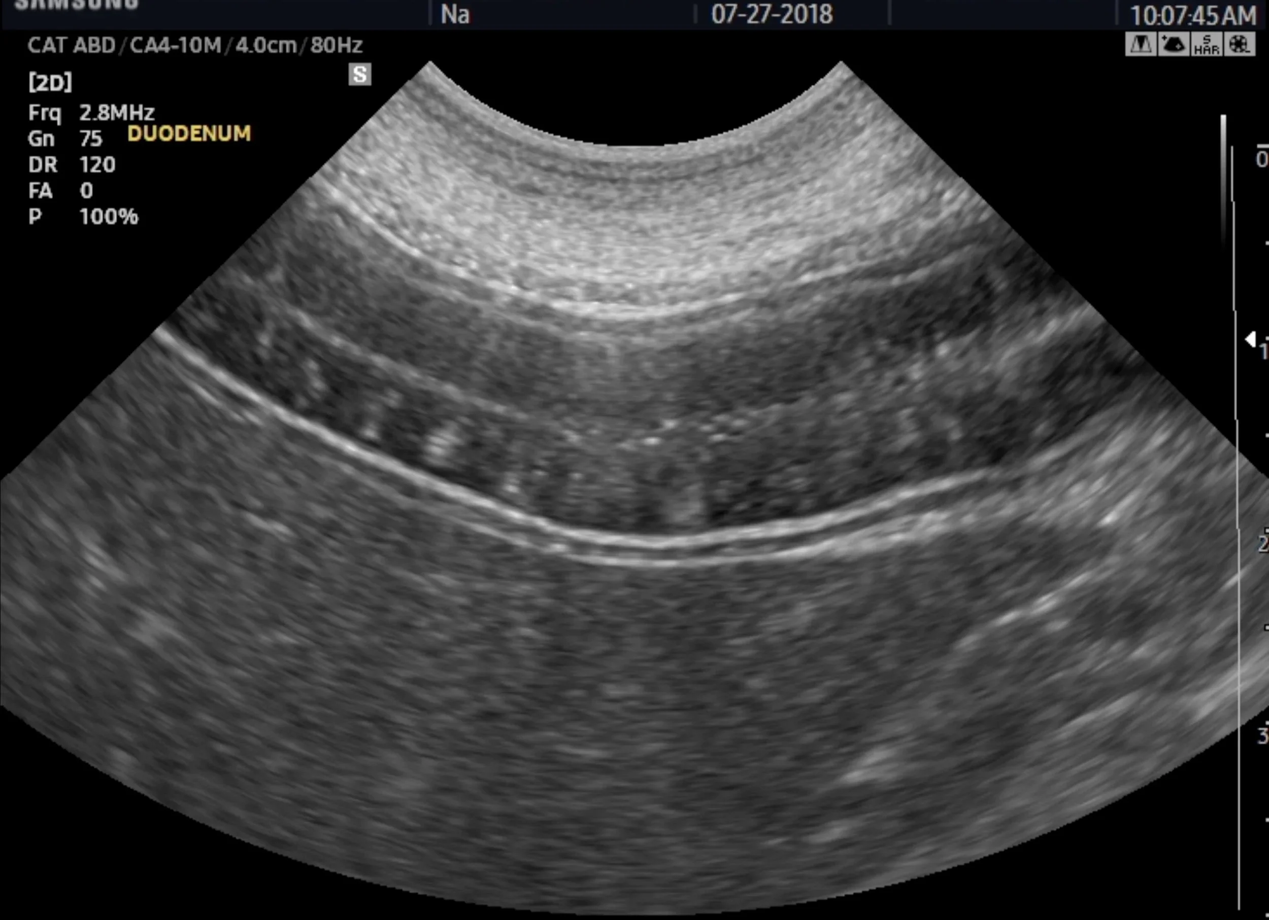
Parsley's duodenum, the first stretch of the small intestine. The bright white speckles scattered through the intestinal wall are swollen lymphatic channels.
Ultrasound lets us look inside the body in real time, without surgery, and study the organs in fine detail.
As our board-certified veterinary radiologist, Dr. Jennifer Johnson-Neitman examined Parsley’s intestines, the answer came into focus: lymphangiectasia. In this condition, the tiny lymphatic channels that help the intestine absorb nutrients become swollen and dilated, so the body struggles to get what it needs from food.
It's a well-known cause of exactly the kind of stubborn, long-running digestive trouble Parsley had been fighting for years.
A Diagnosis Changes Everything
With a clear diagnosis, Parsley's veterinary team could build a treatment plan aimed at the real cause: a low-fat diet and low-dose steroids, plus an internal medicine referral to help her body finally absorb and hold onto what it needed.
Parsley Today
The change in her has been remarkable. Her family reports that Parsley is doing fantastic — her stool is normal, the frightening symptoms are gone, she's put weight back on, and she's bright, comfortable, and herself again.
Perhaps the most telling part came from her own veterinarian, who, knowing how long and how severely Parsley had struggled, admitted to being only fifty-fifty that she'd ever truly improve. Parsley proved the hopeful half right.
Why Imaging Matters
That's the difference the right answer can make. When a beloved pet has been suffering and the cause stays hidden, advanced imaging can finally bring it to light — and give a family their happy, healthy companion back.
Follow Along With Us
Love a good happy ending? Parsley is just one of the interesting cases we see in our reading room. Sign up for our newsletter to follow more stories, learn how imaging helps pets like yours, and stay connected with what we do at Oklahoma Veterinary Diagnostic Imaging.